


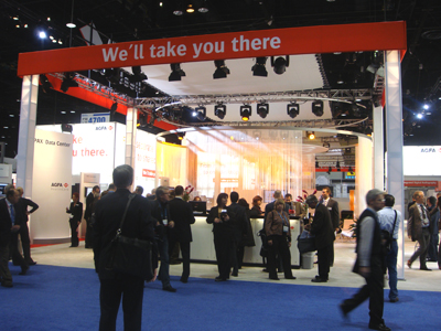
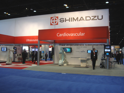
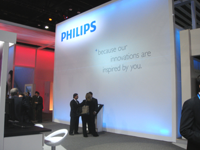
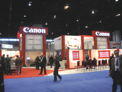
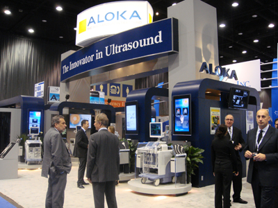
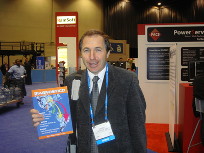
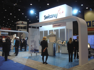
Will Clonts de Pegasus Imaging junto a Ezequiel Domb de Revista Diagnostico George Kovacs de McKesson Posters Electronicos Stand de Nuance Stand de Agfa Stand de Varian Stand de Shimadzu Stand de Philips Stand de Canon Stand de McKesson Stand de Carestream Health Stand de Unfors Stand de Sony Stand de Fujifilm Stand de Hitachi Stand de ICRco Stand de Codonics Stand de Siemens Stand de Aurora Stand de Hologic Stand de Terarecon Stan de Aloka Dr. Daniel Lehrer de CERIM Argentina, lector de Revista Diagnóstico Stand de Swissray Entrada al Hall D del Lakeside Center


With increased integration of refresher course material and scientific papers, RSNA 2008 offers attendees myriad opportunities to not only learn about the latest breakthroughs in the specialty, but also take home knowledge to apply immediately in their work settings.
Medical imaging informatics, which plays a crucial role in the daily practice of radiology, will be featured in scientific and focus sessions, as will quantitative imaging, structured reporting and molecular imaging, said RSNA Scientific Program Committee Chair Robert M. Quencer, M.D., a professor and chair of radiology at the University of Miami School of Medicine.
This year the committee received 10,878 abstracts for consideration 7,052 for scientific presentations and 3,826 for education exhibits. Over the summer, the committee, with its subcommittees, selected 1,803 abstracts as scientific papers and 729 as scientific posters. A separate committee accepted 1,606 abstracts for education exhibits.
Breast Imaging
Defining breast cancer risk and selecting patient-specific screening strategies are developing areas this year, said Jennifer A. Harvey, M.D., subcommittee chair. Presentations examine the cost-effectiveness of MR screening, risk assessment, the role of screening ultrasound and how to address the lower sensitivity of mammography in women with denser breasts.
Other presentations examine digital tomography, breast-specific gamma imaging and MR screening for women with prior breast cancer. There is a marked increase in submitted studies on diffusion- weighted MR, Dr. Harvey said.
Notable trends include the application of cross-sectional and functional imaging using detectors specifically designed for the breast. This includes positron emission mammography, breast specific gamma imaging, CT, tomosynthesis and whole breast ultrasound, said Dr. Harvey. These developing applications may play a role in improving diagnostic capabilities and provide ancillary screening for women at high risk of developing breast cancer.
Cardiac Radiology
Overall, there is strong interest in cardiac CT, with presentations focusing on radiation dose and technique development, said subcommittee chair Andre J. Duerinckx, M.D., Ph.D.
Cardiac subcommittee members identified many great abstracts in two key areas early population studies about clinical acceptance and applications of cardiac CT as well as the use of dualsource CT, said Dr. Duerinckx.
Other sessions will cover radiation safety, cardiac CT technique improvements, plaque imaging, quantitative cardiac radiology and comparisons with echocardiography and nuclear stress testing, he said.
Chest Radiology
In the chest subspecialty, there is continued interest in pulmonary emboli and nodules, said H. Page McAdams, M.D., subcommittee chair.
Pulmonary embolism presentations will explore dose issues and applications in pregnancy, while studies in nodule classification will examine computer-aided detection (CAD), nodule volumes and screening. Many abstracts focus on CAD applications beyond mere nodule definition, Dr. McAdams said.
Other remarkable topics are texturebased classification of interstitial pneumonia, variability in pulmonary nodule volume software and dual-energy CT of the peripheral vessels, said Dr. McAdams.
Emergency Radiology
Subcommittee Chair Diego B. Nuñez Jr., M.D., M.P.H., noted continuing trends toward validation of current practices, such as CT utilization and radiation safety in the emergency setting. He observed particular interest in multidetector CT for abdominal trauma, particularly as it relates to scanning protocols, delayed imaging and multiplanar display.
Notable presentations will examine cumulative data on modality utilization, the appropriateness of Prospective Investigation of Pulmonary Embolism Diagnosis (PIOPED) II criteria for patients aged 40 years and younger, the importance of delayed CT in blunt trauma and pelvic CT angiography in blunt trauma using 64-slice multidetector CT, said Dr. Nuñez.
Gastrointestinal Radiology
CT colonography as a viable screening study for polyp detection remains a popular topic, said Erik K. Paulson, M.D., subcommittee chair. There is also continued interest in exploring the efficacy of this technology, applications of computer-aided detection and development of novel bowel preparations, he said. He also noted increased interest in analyzing the biologic behavior and treatment response of tumors with quantitative CT and MR techniques, including 3T MR applications.
Dual-energy CT is now offered on clinical scanners and the first wave of critical assessment of this technology will be presented, Dr. Paulson said.
Researchers in the subspecialty also continue to explore novel methods to reduce radiation dose without suffering loss in diagnostic accuracy.
Genitourinary Radiology
Marcia C. Javitt, M.D., subcommittee chair, reported more studies on diffusion- weighted imaging of masses in the kidneys, ovaries and prostate. New data also were submitted reporting long-term follow-up on patients who underwent renal tumor and fibroid ablation, as well as cryotherapy.
A groundbreaking area of study, said Dr. Javitt, is the linkage of gene expression to tissue characterization. In these studies, she said, the morphology visible on cross-sectional imaging studies was compared with genetic markers of renal cell cancer. She noted another practical study of contrast-induced nephropathy and nephrogenic systemic fibrosis, which reported on the use of glomerular filtration rate measurement techniques.
Health Services Policy and Research
A broader range of research topics and some novel submissions will be part of the program this year, said Subcommittee Chair Ruth C. Carlos, M.D., M.S. Notable topics include disparity in types of imaging related to socioeconomic status or insurance coverage, the effects of including a patient photo with a radiologic exam, occupational stress in radiologists and evaluation of an outpatient imaging center where radiologists consult directly with patients, said Dr. Carlos.
Other presentations this year will address resident education, evidence-based medicine and guideline development.
Informatics
This years submissions indicate that business intelligence and data analytics are being increasingly used for clinical and operational data mining, said Keith Dreyer, D.O., Ph.D., subcommittee chair. He also noted a significant trend in the understanding of natural-language for radiology reports.
Informatics presentations will cover topics such as the effects of structured reporting and decision support, using the iGoogle dashboard to monitor a radiology department, the RAD-DASH Web-based graphical business platform for managing radiology performance, quality metrics and the National Cancer Institutes Cancer Biomedical Informatics Grid (CaBIG), said Dr. Dreyer.
Molecular Imaging
Subcommittee Chair Umar Mahmood, M.D., Ph.D., noted an increased variety of applications and agents in this years submissions.
This is exactly what we want as the field moves more into the clinic, he said.
Topics of note include new MR molecular imaging methods, multimodal agents, the use of fluorodeoxyglucose PET to predict cardiovascular risk, a new PET tracer to assess innervation and increased use of MR smart agents. Human application represents a small minority of abstracts, but I think it has increased somewhat compared to previous years, said Dr. Mahmood.
Musculoskeletal Radiology
Presentations this year represent more attempts to characterize bone and soft tissue tumors using advanced MR techniques like diffusion imaging and spectroscopy, said David A. Rubin, M.D., subcommittee chair. More emphasis is placed on quantitative analysis of images and data, as opposed to qualitative analysis, Dr. Rubin added.
This trend is present in tumor characterization, imaging methods for osteoporosis and even tendon sonography, he said.
Dr. Rubin also noted a trend toward using CT and ultrasound guidance, rather than fluoroscopy, to increase the safety of risky procedures like cervical spine injections. Other presentations, he said, will explore applications of diffusion tensor imaging and tractography for the imaging of peripheral nerves and muscles, use of diffusion MR to help distinguish benign from malignant musculoskeletal tumors, ultrashort TE MR applications, percutaneous tumor and pain management techniques and tendon sonoelasticity.
Neuroradiology/Head and Neck
Utilization of advanced imaging techniques like diffusion-weighted imaging, perfusion with MR and CT and spectroscopy for all masses in the neck are increasing, said Subcommittee Chair Mauricio Castillo, M.D. He noted that sonographic elastography for diagnosis of neck masses is also beginning to emerge as a mature clinical imaging technique.
Diffusion-weighted imaging of the spinal cord is being refined and beginning to be clinically utilized and helpful, said Dr. Castillo.
Diffusion-weighted imaging of the intervertebral disc may be able to identify an abnormal disc before other techniques. Also, the effects of iron deposition in the brain and its relationship to several neurodegenerative disorders are being investigated with susceptibility weighted imaging.
Also presented this year will be several techniques that may help to distinguish between symptomatic and asymptomatic atherosclerotic plaques in the internal carotid arteries, said Dr. Castillo.
Nuclear Medicine
PET imaging techniques with respiratory gating and new hybrid technology such as gamma ultrasound will be highlighted this year, said Milton J. Guiberteau, M.D., subcommittee chair.
Presentations will also represent the emerging literature of correlative clinical imaging technologies such as fluorodeoxyglucose (FDG) PET, CT and MR, Dr. Guiberteau continued.
In addition to using nuclear medicine to assess and predict lesion response to more established conventional treatments, important topics will include nuclear medicine evaluation of the success of emerging therapies, such as liposomal-mediated oncologic treatments and interventional therapy outcomes.
A nuclear medicine quantitative imaging session will address FDG-PET standardized uptake value (SUV) determinations in a variety of clinical settings, Dr. Guiberteau said.
Pediatric Radiology
Subcommittee Chair Lane F. Donnelly, M.D., said attendees can look forward to important presentations on CT dose, helical MR, diffusion tensor imaging for body applications and pediatric interventional radiology.
Physics
Notable physics abstracts explore noncontrast MR detection of tumor microvasculature and a four-view CAD system for breast masses, said subcommittee chair Martin J. Yaffe, Ph.D. Other presentations will address distortion-free MR imaging near metallic implants, prognosis of breast carcinoma using computer extracted morphological and kinetic features in dynamic contrast-enhanced MR and monitoring intrahepatic and subcutaneous hepatocellular carcinoma growth with 23Na and 1H MR.
Radiation Oncology/Radiobiology
Subcommittee chair James S. Welsh, M.D., M.S., said notable topics this year include consolidative radiotherapy for non-Hodgkin lymphoma, hepatic arterial embolization with Holmium-166 poly (L-lactic acid) to predict distribution of therapeutic dose and immunomodulator therapy to enhance radiotherapy response. There are also a variety of interesting radiation biology subjects, said Dr. Welsh, along with strong submissions for the Bolstering Oncoradiologic and Oncoradiotherapeutic Skills for Tomorrow (BOOST) program.
Vascular and Interventional Radiology
MR-guided galvanotherapy for prostate cancer, cryotherapy for painful bone metastases, drug-coated percutaneous transluminal angioplasty balloons to improve patency and percutaneous recanalization after failed surgery are remarkable topics this year, said John A. Kaufman, M.D., subcommittee chair. Oncologic interventions constitute the single largest topic, he said.
Global Commitment to Quality
The substantial input into the quality of the meeting by radiologists and scientists involved in imaging research throughout the world will make RSNA 2008 an important meeting for all those involved in radiology and its allied fields, said Dr. Quencer.